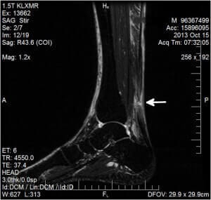
After I had my first ever MRI in October 2013 in order to check whether I had fully ruptured my achilles tendon, I asked the imaging center for a copy. They gave me around 20 different images on a CD, and I have pasted the most relevant of these below. Note that my MRI cost around $1,500, and I was responsible for around $650 of that after insurance covered the rest. I have since considered starting my own imaging center as it seems like the easiest way in the world to get rich, but am lacking the motivation and start-up capital for now:-(
For more details, see my achilles tendon rupture diary.
I supposedly had a 1.2 centimeter (cm) long full rupture of my achilles tendon, 7 cm above the calcaneal (heel) insertion of the tendon. Interestingly, most achilles tendon ruptures occur between 2 cm to 6 cm above the calcaneal insertion (an area called the “watershed zone”) where the blood supply is poorest. According to some experts, ruptures higher than this heal faster due to a superior blood supply.
In fact, when an achilles tendon rupture is too high and close to the calf muscle, even pro-surgery American doctors usually recommend non-surgical conservative treatment. It is not a good idea to try to stitch tendon to muscle. I have read a number of blogs where people with very high ruptures near the calf muscle healed really well via non-surgical treatment. In my case, since the rupture was just 1 centimeter above the watershed zone, my doctor felt that was a good thing insofar as healing potential and speed of healing went.
Among the various MRI images of my lower leg, several supposedly showed some pre-existing tendinosis, which is quite common in those who rupture their achilles tendon. My doctor said that the blood flow around my lower legs was good. One day I will try to make a radiologist friend who can show me where on my numerous MRI images I can see the pre-existing tendinosis and good blood flow.
Prior to my sudden rupture, I had never suffered from any kind of tendon pain to indicate any issues with the tendon such as tendinosis, and this is also quite common for most people who have this injury (i.e., there is no indication whatsoever that you are at risk of suffering from it).
In the MRI image at the top of this page, the exact area of rupture is depicted via a white arrow. An intact achilles tendon is usually represented by a long thin sold black strip. Any white area in place of solid black within that strip is usually a sign of a rupture. It is a bit unusual that despite my only getting the MRI done 10 days after rupture, the gap in tendon edges was only 1.2 cm long. During those 10 days, I was very active and walking (albeit with a slight limp), driving and even climbing and coming down stairs on several occasions. I was lucky that the gap did not widen. You can click on the image to enlarge it.